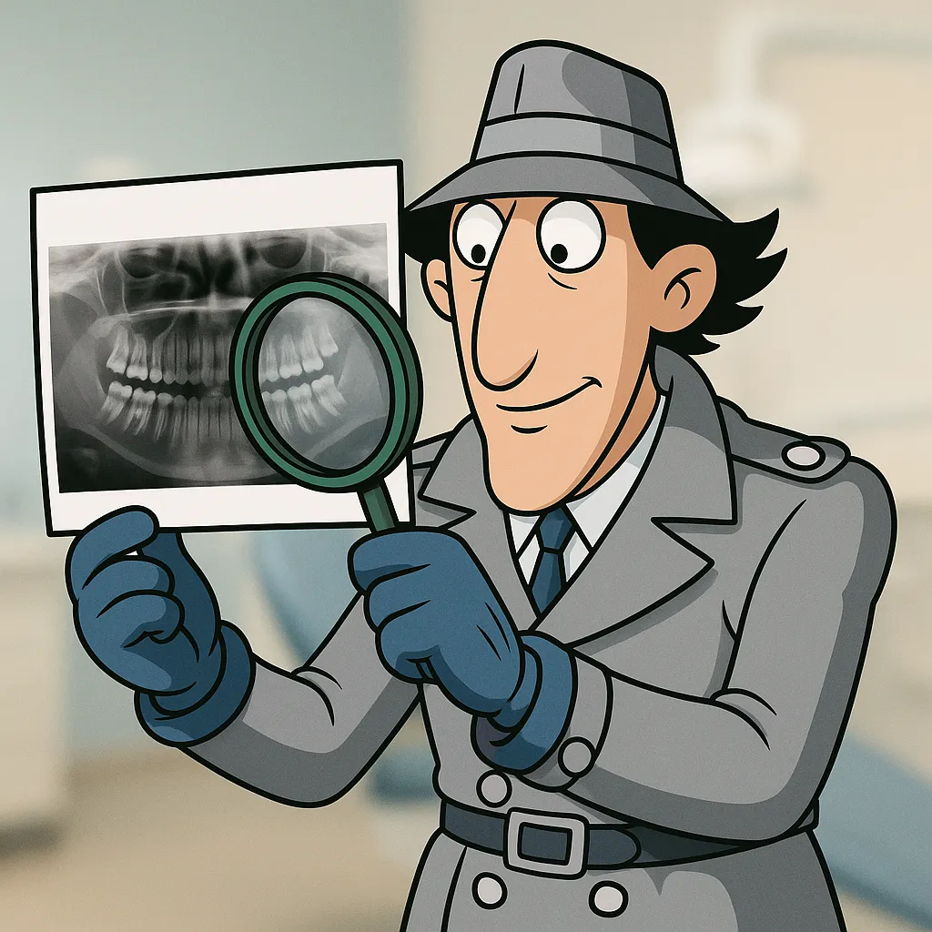
Reading X-rays Like a Super-Detective
How AI Diagnostic Imaging Is Revolutionizing Dental X-rays
(And What It Still Can’t Do)
Let’s face it—interpreting dental X-rays isn’t always black and white. Yes, there are obvious signs of decay, fractures, and bone loss. But then there are the close calls. The shadowy interproximal areas. The early-stage lesions you think you see. The periapical change that doesn’t quite scream pathology but nags at your gut.
This is where many dental professionals are now finding an unlikely ally: artificial intelligence.
Diagnostic Uncertainty and Time Pressure
No dentist wants to miss something. But in a real-world clinical setting—where you’re moving from operatory to operatory, balancing hygiene checks, emergencies, and consults—it’s impossible to stay 100% sharp, 100% of the time.
You’re making dozens of diagnostic calls every day. And those calls often hinge on interpreting grayscale images under suboptimal conditions. Combine that with patients who expect definitive answers, and the pressure builds fast.
And while seasoned dentists may develop strong diagnostic instincts over time, even the best clinicians will admit it: fatigue, distractions, or subtle radiographic signs can lead to oversight.
Gaps in Consistency, Patient Trust, and Legal Risk
Here’s where things can go wrong. A missed lesion that turns into a root canal. A fracture that goes undetected until the tooth splits. A patient who questions your recommendation because they “don’t see anything” on the screen.
That’s not just a clinical problem—it’s a reputational one.
And it’s not just about missing the diagnosis—it’s about explaining it. Patients today are more informed (and skeptical) than ever. If they don’t see the issue, they may not accept the treatment plan. That creates friction and delays care.
It also opens up legal exposure. Inconsistent documentation and missed early signs can come back months—or even years—later.
What AI Can Do in Diagnostic Imaging
AI in dental imaging isn’t science fiction. It’s happening now—and it’s changing the way practices operate.
Let’s break down what today’s AI software can realistically do:
Detect early-stage caries in enamel and dentin, especially in posterior interproximal regions where visibility is limited.
Highlight periapical radiolucencies, helping identify potential abscesses or granulomas before symptoms escalate.
Flag bone loss patterns consistent with periodontal disease progression.
Track changes over time, comparing new and previous images to help monitor lesion development.
Generate visual overlays, making it easier for patients to understand and trust your recommendations.
These tools don’t replace your expertise—they enhance it. Think of it like a diagnostic assistant who’s always alert, always consistent, and able to catch what might slip by in the rush of the day.
AI can also serve as a training aid for newer clinicians, helping them build pattern recognition and confidence. It offers structure, support, and a second set of eyes that never miss a detail.
But Let’s Be Clear of What AI Can’t Do
It’s not magic. It’s still a tool—and it has limits.
AI can’t diagnose on its own. It highlights, not decides.
It doesn’t understand patient history, context, or clinical presentation. A flagged spot might be an artifact or an old scar.
It won’t replace your judgment. If anything, it requires even more of it—because now you’re not just interpreting the image, you’re interpreting the software’s analysis too.
Smart clinicians use AI as a confirmation, not a crutch.
Real-World Impact: What This Means for Your Practice
You might be wondering—does this actually improve outcomes?
Here’s what practices integrating AI imaging are seeing:
Higher patient case acceptance. When you can show patients what the software sees—highlighted in color, annotated, and backed by data—they say yes faster.
Improved accuracy. Especially with early-stage lesions that might otherwise be overlooked.
Increased confidence. For both younger associates and seasoned providers who want a double-check on borderline cases.
Better documentation. AI reports often come with annotated image capture, making records clearer and legally stronger.
In other words: less second-guessing, more certainty, and smoother communication.
So, Should You Adopt AI Imaging?
If you’re committed to diagnostic excellence, it’s worth exploring. Many AI dental software platforms integrate seamlessly with your current imaging system. There’s a learning curve, sure—but nothing compared to the potential gain in precision, protection, and patient trust.
We’re not saying AI will solve everything. But if it helps you catch one more fracture a week… prevent one more unnecessary root canal… or help one more patient say yes with confidence—then it’s already doing its job.
